

Science & Health Communication
Translating complex science into accessible communication across healthcare, education, and public outreach.
I specialize at the intersection of science, design, and communication, helping organizations make information clear and engaging for diverse audiences. My work spans web, digital media, visual storytelling, educational design, and public-facing communication.
Case Studies
PocketHealth’s Report Reader
March, 2025 | Toronto, Ontario


Following completion of my MSc in Biomedical Communications at the University of Toronto, I joined PocketHealth as the company’s first Biomedical Communicator.
Working closely with designers, engineers, and leadership, I developed over 300 patient-facing medical visuals and helped shape the visual language of PocketHealth’s report reader experience, focusing on improving patient understanding through accessible digital healthcare communication.
The work highlighted how visual communication can reduce ambiguity and improve patient understanding in radiology.
“What impressed me most was Brendan’s curiosity about how systems worked under the hood. He brought together anatomical expertise, illustration, and technical thinking to create meaningful patient-facing solutions.”
— Sai Siddulugari, Engineer at PocketHealth
Making Complex Reports Simple
Full case study and process documentation.

Radiology reports are often difficult for patients to interpret independently without visual context.
“Interactive Body Atlas” - B.Lazar & PocketHealth Engineering Team | February, 2025
This project was created over a few days and was the first time all of PocketHealth’s application features could be used in the same place. It combined report reader, organ tagging on scans, and accurate medical visuals to help educate and inform patient’s through an engaging and interactive way. View the case study for the full demo.
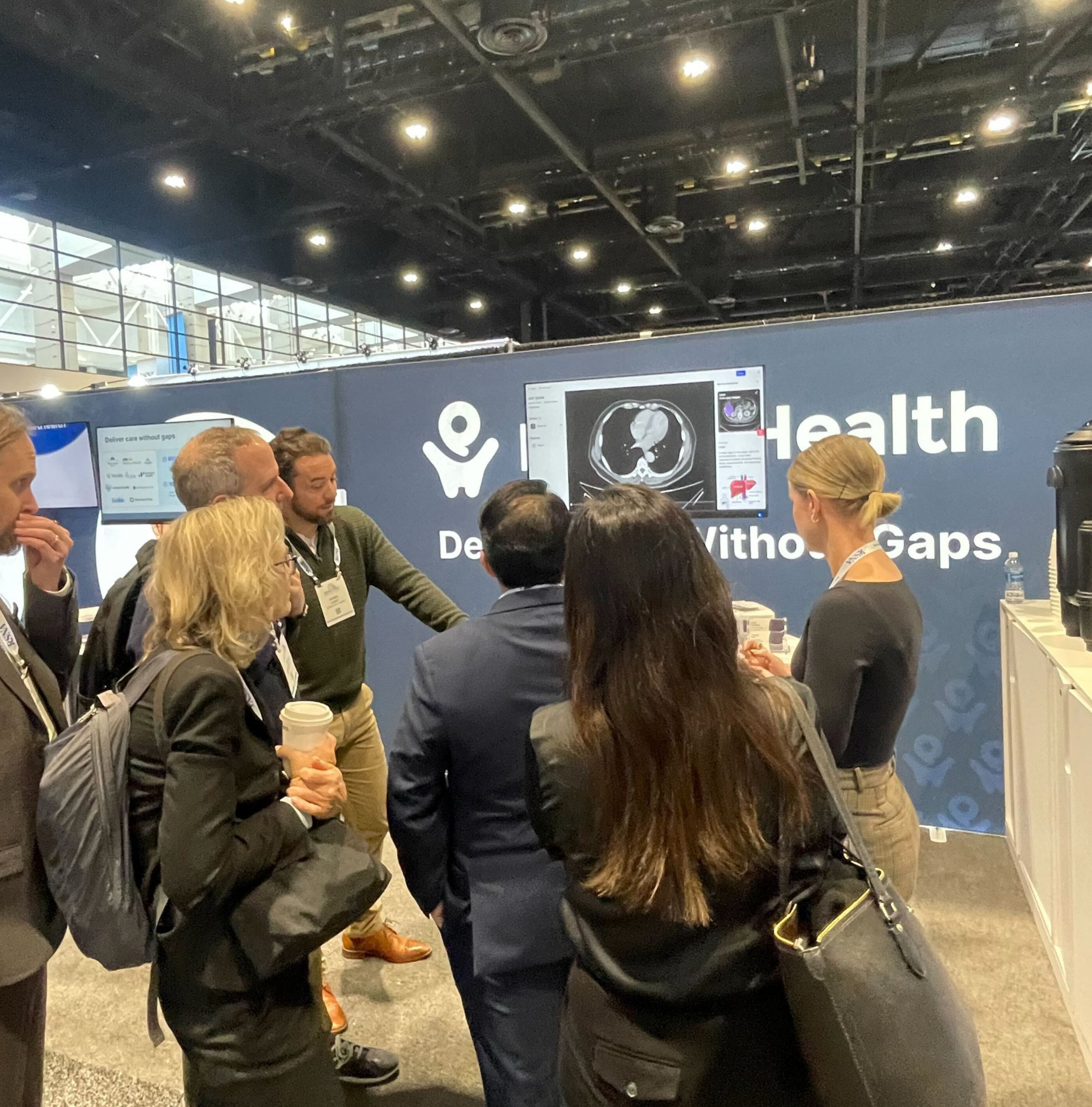
PocketHealth team demonstrating the Report Reader platform at a radiology conference.
The project focused on simplifying complex imaging concepts into clear, accessible educational systems for a patient-audience that could integrate directly into PocketHealth’s digital platform.
The Canada Prize for Earth Science
November, 2025 | The Canada Prize Foundation, Calgary Alberta

The Canada Prize for Earth Science is a developing international award intended to recognize transformative contributions in Earth Science.
This project reimagined the Prize’s visual identity and digital presence to communicate scientific prestige, institutional credibility, and contemporary accessibility.
This project explored how branding and digital communication can establish legitimacy and public trust for emerging scientific institutions.
Communication Challenge
Determining how a newly developing scientific prize could visually position itself alongside internationally recognized awards such as the Nobel Prize, Japan Prize, and Kavli Prize while maintaining a distinctly Canadian identity was the central challenge.
The work balanced institutional formality with modern accessibility while creating a visual system flexible enough for web, outreach, and future international growth.
The process involved extensive stakeholder coordination and iterative refinement around symbolism, accessibility, and Canadian identity.
Full case study and process documentation coming soon.
Neuroscience Self Portrait
October, 2023 | MSc Biomedical Communications, University of Toronto
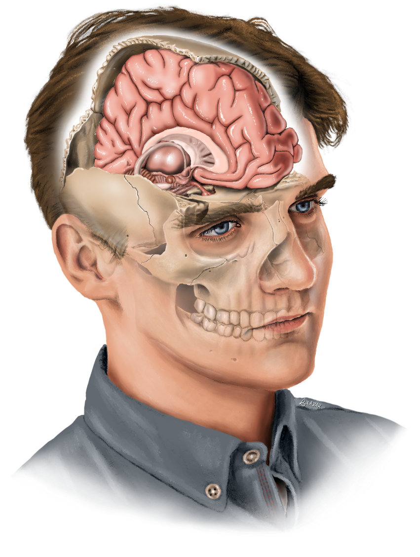
This self-portrait combined portraiture, neuroanatomy, and 3D anatomical reference workflows to explore the intersection of identity and medical visualization.
“It was important to me to make it as accurate as possible; the angle, view, anatomy, and placement of all structures is exactly how you would see it if you could look into someone’s mind at this angle. I also decided to draw the skull cutaway along the suture lines, as opposed to straight cuts - every bone in the skull here is on its own layer.”
Developed through extensive anatomical referencing, including custom skeletal photography, 3D maquettes in Autodesk Maya, and iterative expert feedback to ensure structural accuracy.
This project explored how scientific illustration can communicate both technical anatomy and personal narrative.
Full case study and process documentation.
Research & Process Work
“An Animation on Retinal Organoid Chimeras for Studying Inherited Retinal Diseases”

Toronto, Ontario | August, 2024
Inherited retinal diseases opening sequence - B.Lazar, 2024.
To visualize retinitis pigmentosa, I modelled and animated an eye. The outro transition mimics the tunnel vision patients gradually experience with this incurable disease, highlighting the real-world impact of inherited retinal diseases and the importance of ongoing retinal research.
From 2023 to 2024, I led the development of an animation exploring retinal organoid chimeras and their potential applications in inherited retinal disease research. This thesis project was created in collaboration with researchers at the Ballios Lab at Toronto Western Hospital and faculty at the Biomedical Communications program at the University of Toronto. The process combined stakeholder meetings and consults, scientific research, visual storytelling, script writing, storyboarding, and animation development to communicate a complex emerging and promising area of biomedical science to broader audiences.
The project also involved regular lab visits, visual reference gathering, and close collaboration with researchers to ensure client goals in addition to scientific and visual accuracy throughout development.

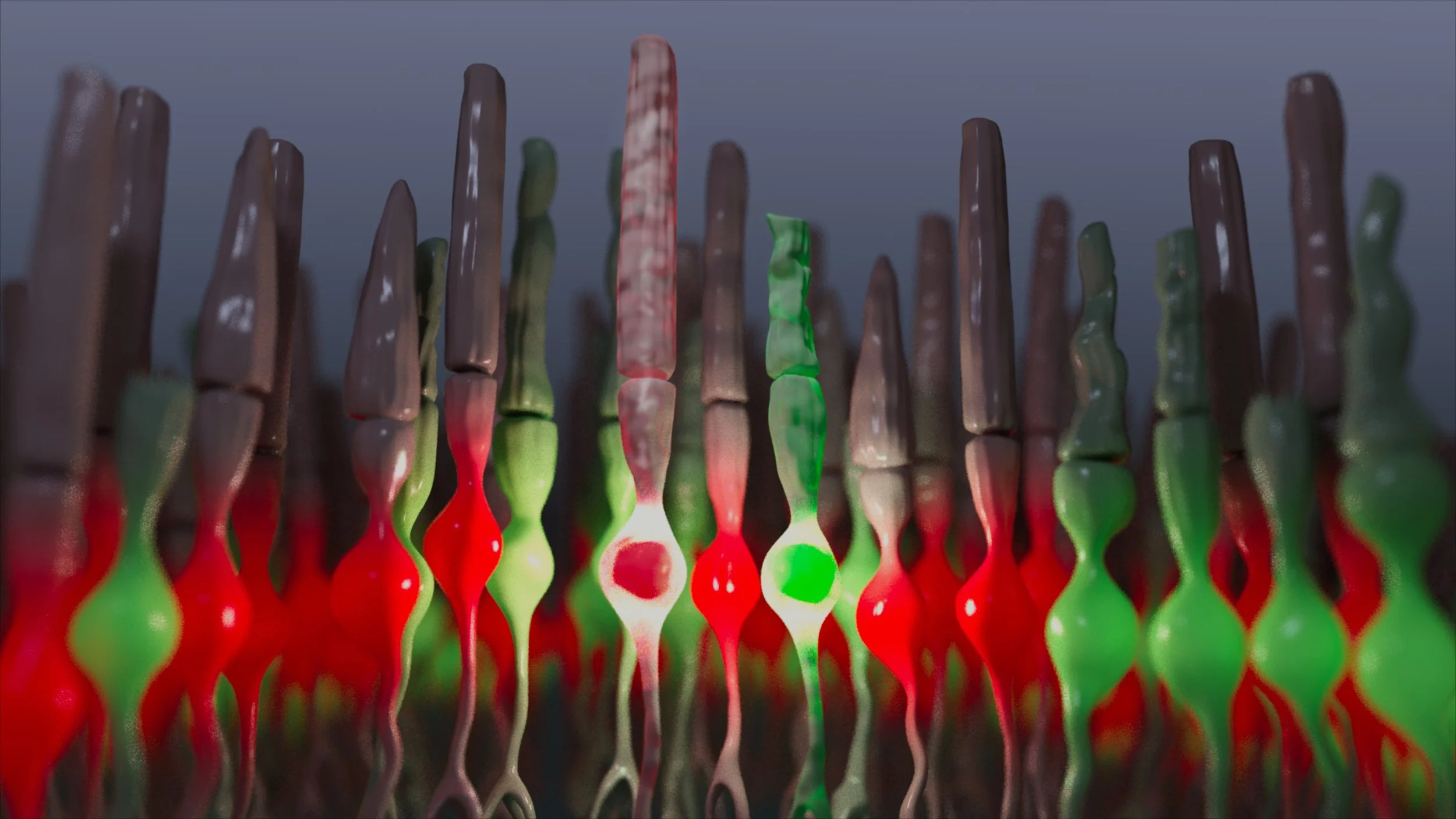
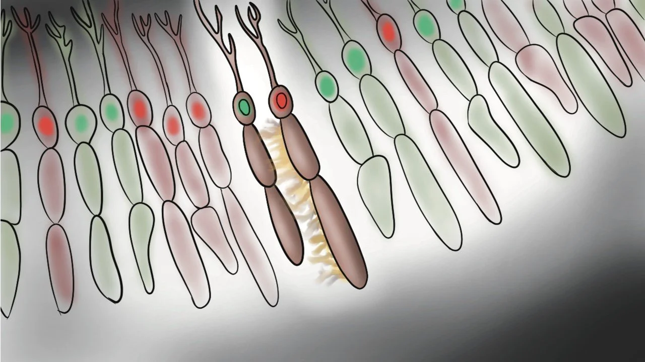
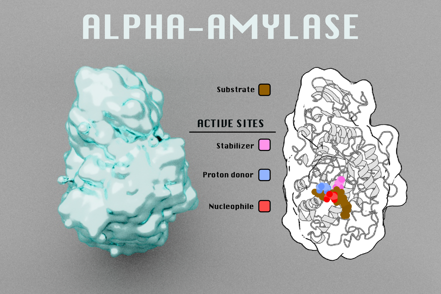
Concept render of scientifically accurate retinal chimeroid photoreceptors - B.Lazar, 2024
These visuals were developed from sketches provided by researchers in the Ballios Lab exploring potential chimeroid photoreceptor structures. To support the broader communication goals of the animation, the designs were adapted toward more recognizable classical photoreceptors so the visuals would remain accessible to patients, students, and conference audiences. Red cells are healthy, while green cells are diseased, based on real fluorescent tagging of cell nuclei used in lab. Rendered in Autodesk Maya.
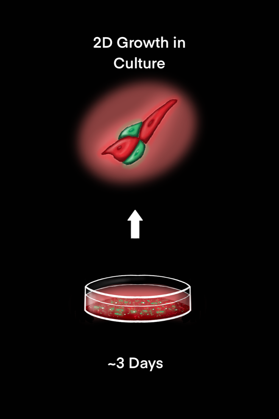
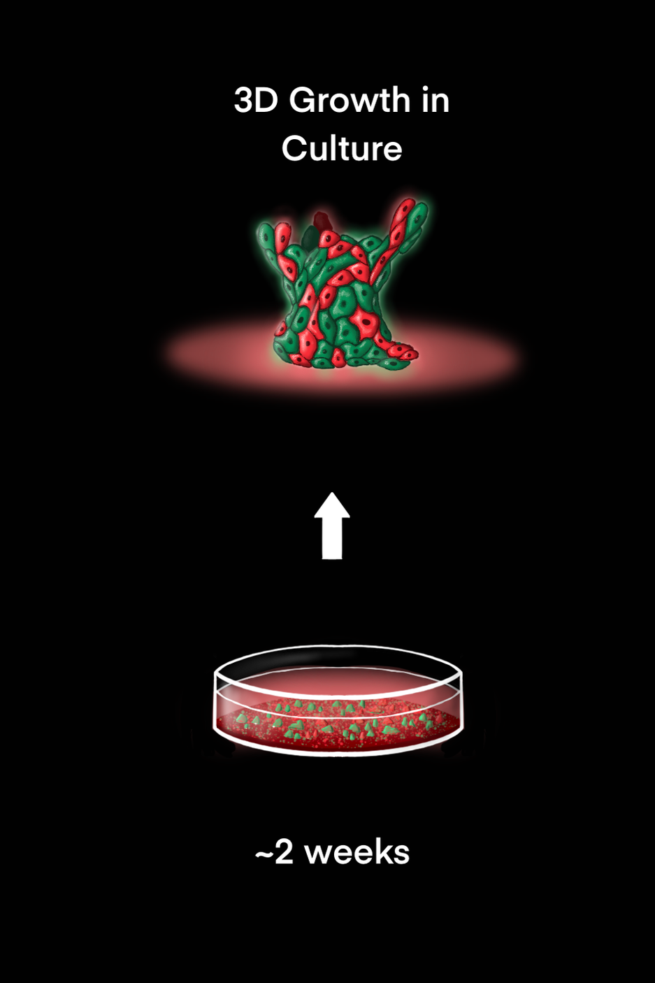
Early stage concept art of 2D and 3D growth of chimeroids in cell plates - B.Lazar, October 2023
These early concept pieces helped test how the two main growth stages could be visualized before being developed into the final animation.
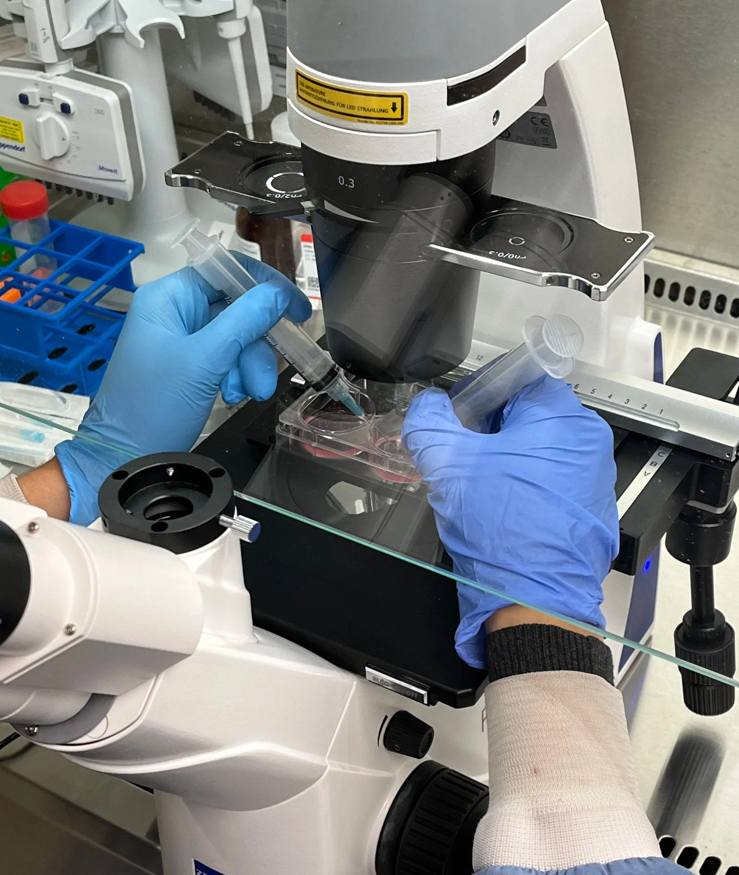
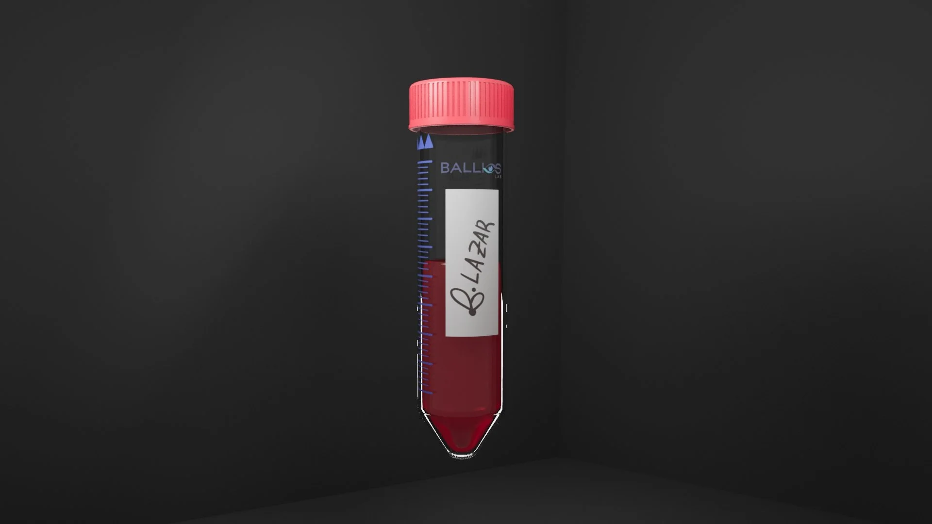
Photo of retinal organoid dissection being done in the lab - taken by B.Lazar with permission from the Ballios Lab, used for research and documentation purposes
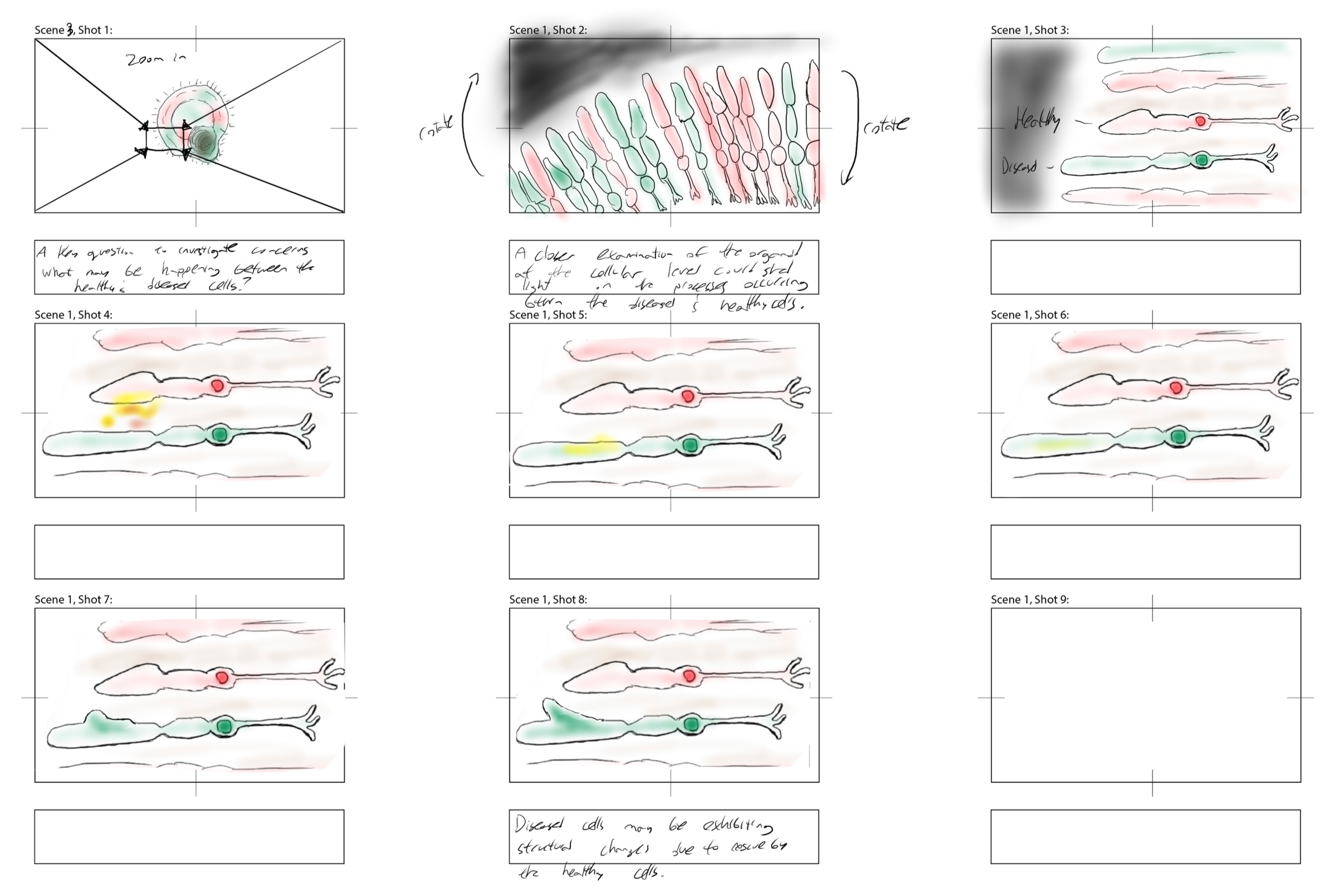
Early Storyboarding (left) and Animatic Frame (right) of Hypothetical Photoreceptor Interactions - B.Lazar, October & December 2023
The central theory behind their work was that healthy cells were “talking” to diseased cells, transferring material to effectively rescue them from the fate of cell death imbued in their genetic code. This is not observable, but measured based on rates of cells surviving. And it was a very fun challenge to understand how to represent this in an animation.
Hypothetical photoreceptor interactions within an organoid - B.Lazar, July 2024
Final animated sequence, including transition between sectioned organoid and microscopic view of photoreceptor talk. Rendered with Maya, edited with After Effects, compiled into larger animation with Premiere Pro.
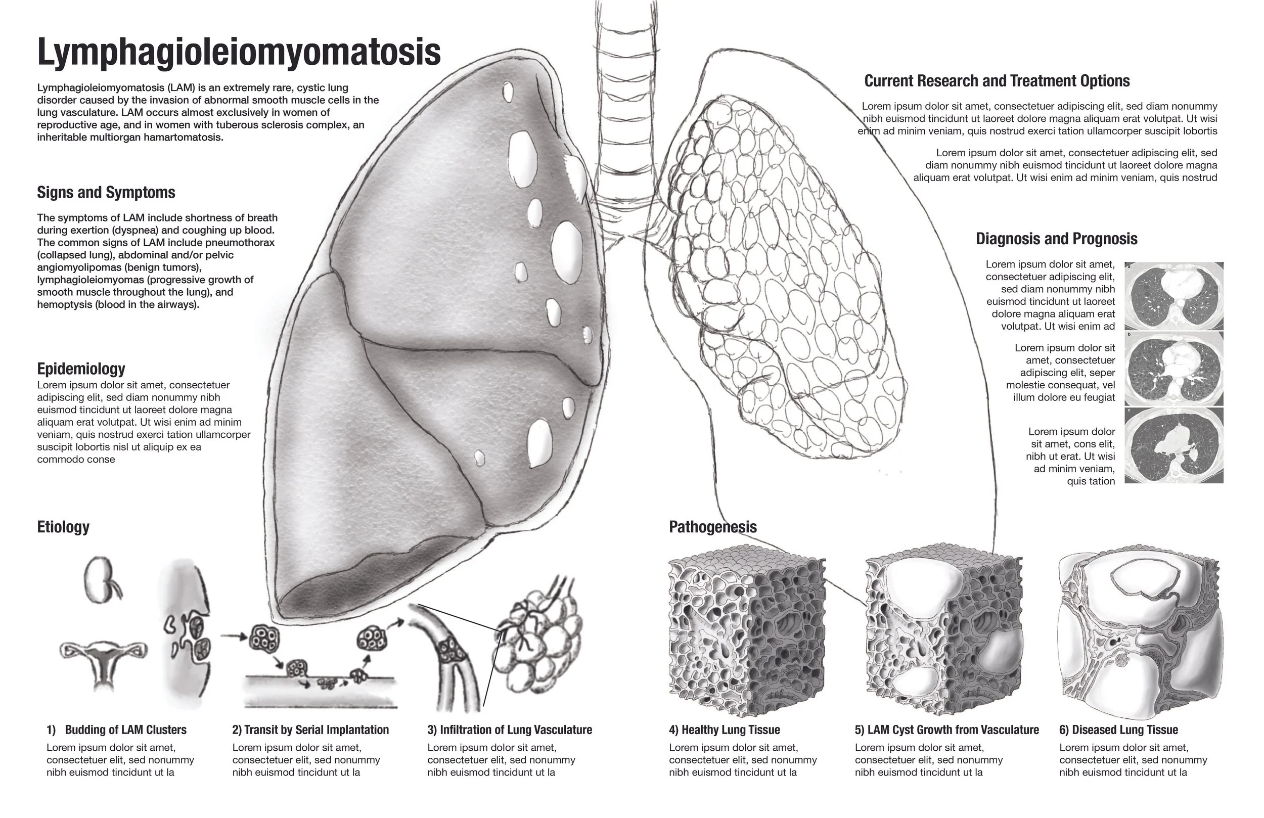
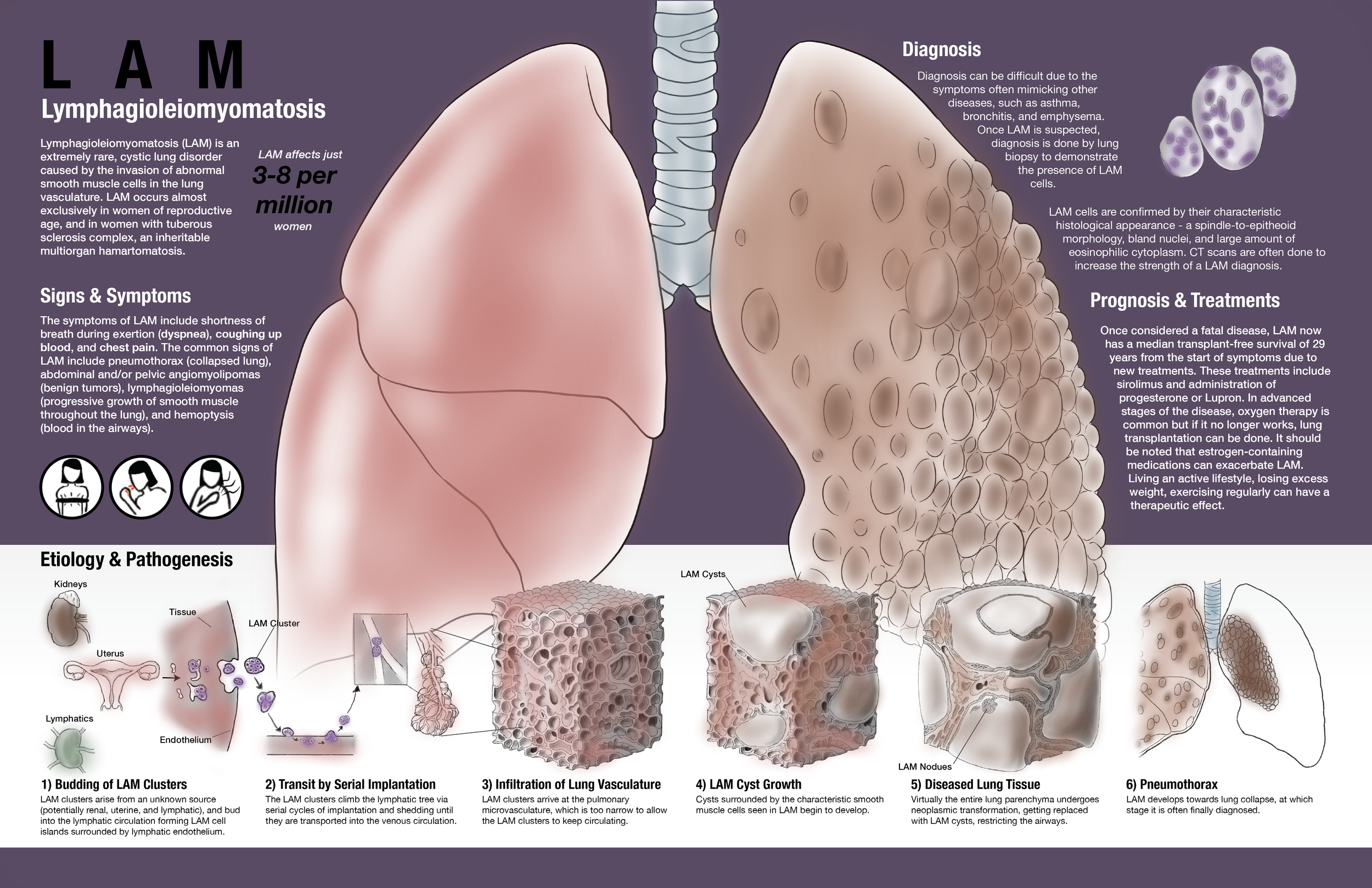
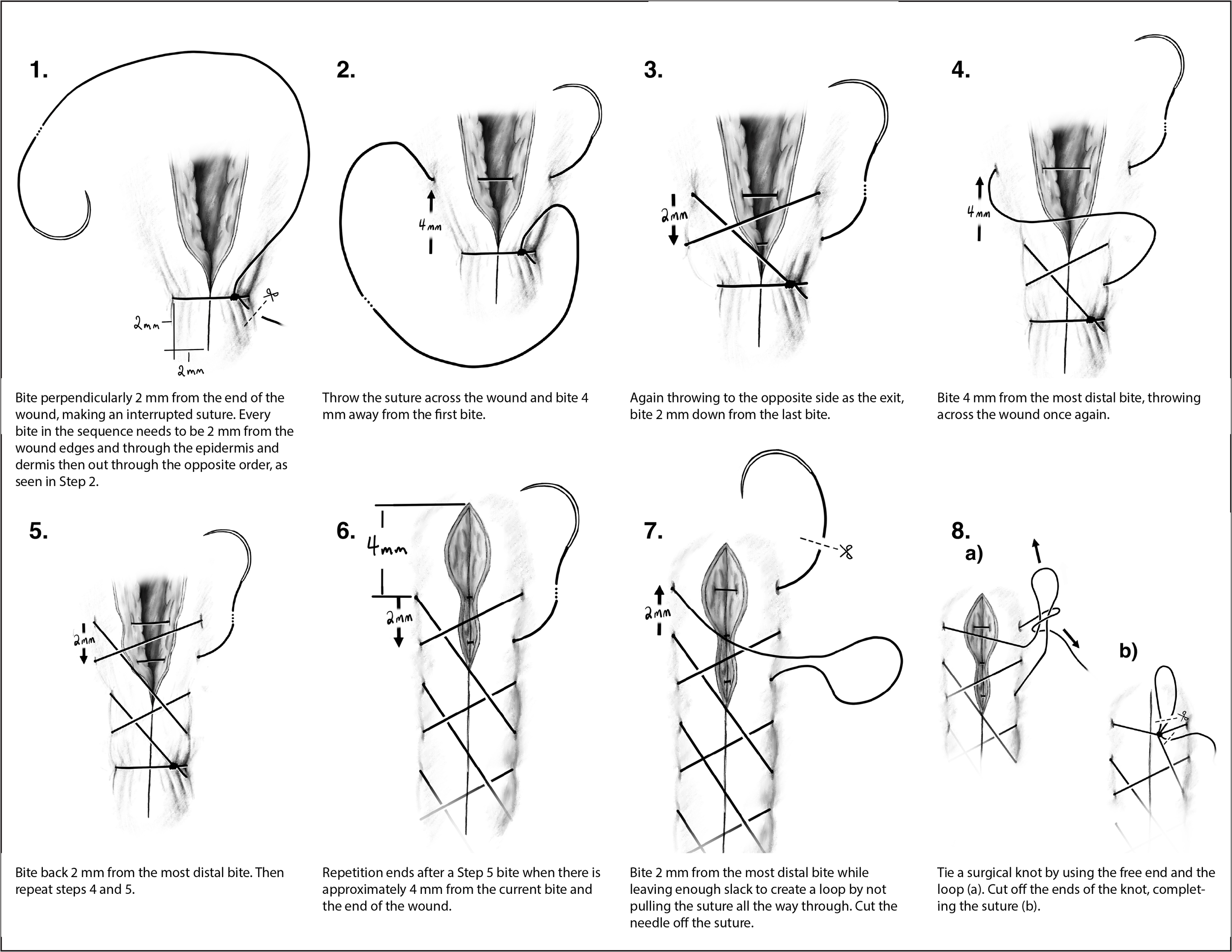
Lymphangioleiomyomatosis (LAM) Informational Spread
View section on desktop for full process work.
April, 2024 | MSc Biomedical Communications, University of Toronto
(Revisions & additional process work on this project coming soon)
Process work

Displayed at the 2024 Association of Medical Illustrators Conference in Rochester, New York.
View section on desktop for full process work.
Draft work - Final Sketch (left) & Coloured Thumbnail (right) of more Developed Sketch - B. Lazar, 2024.
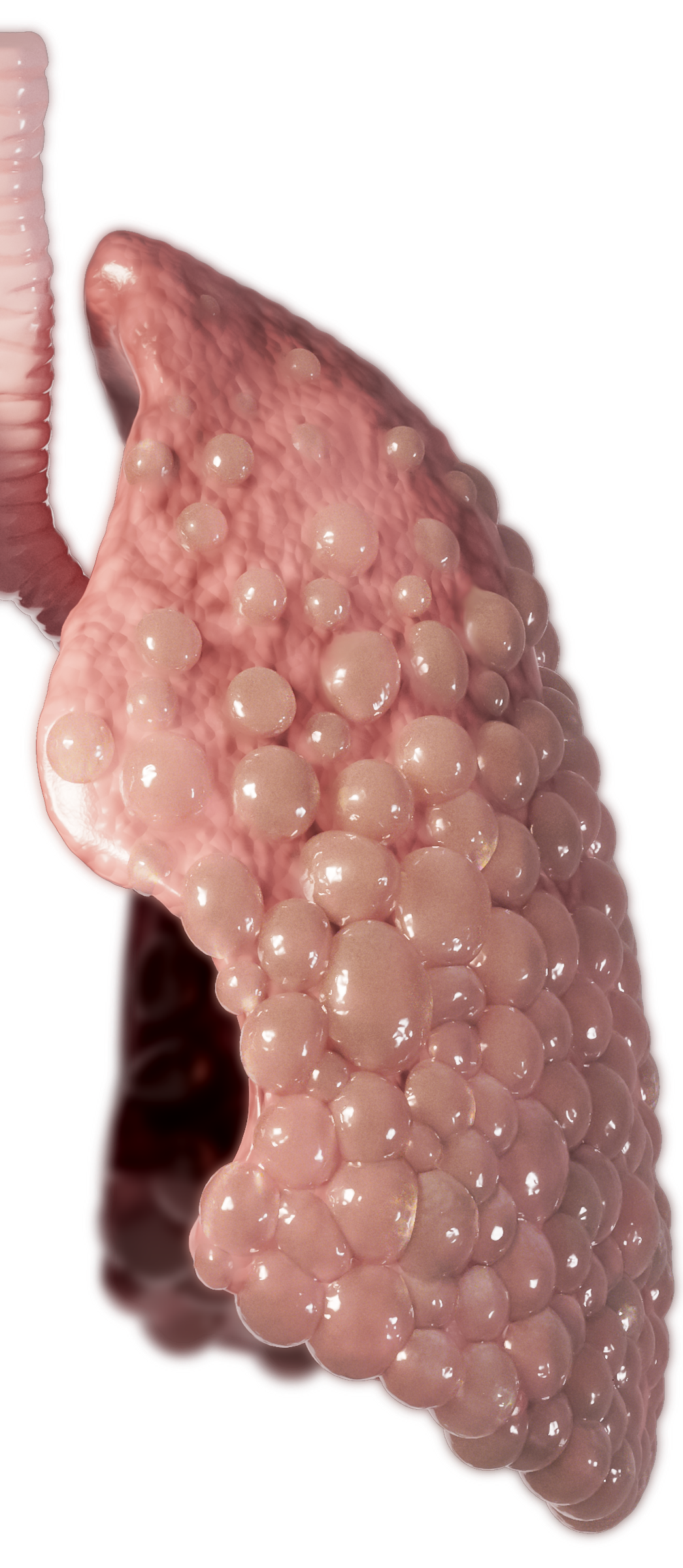
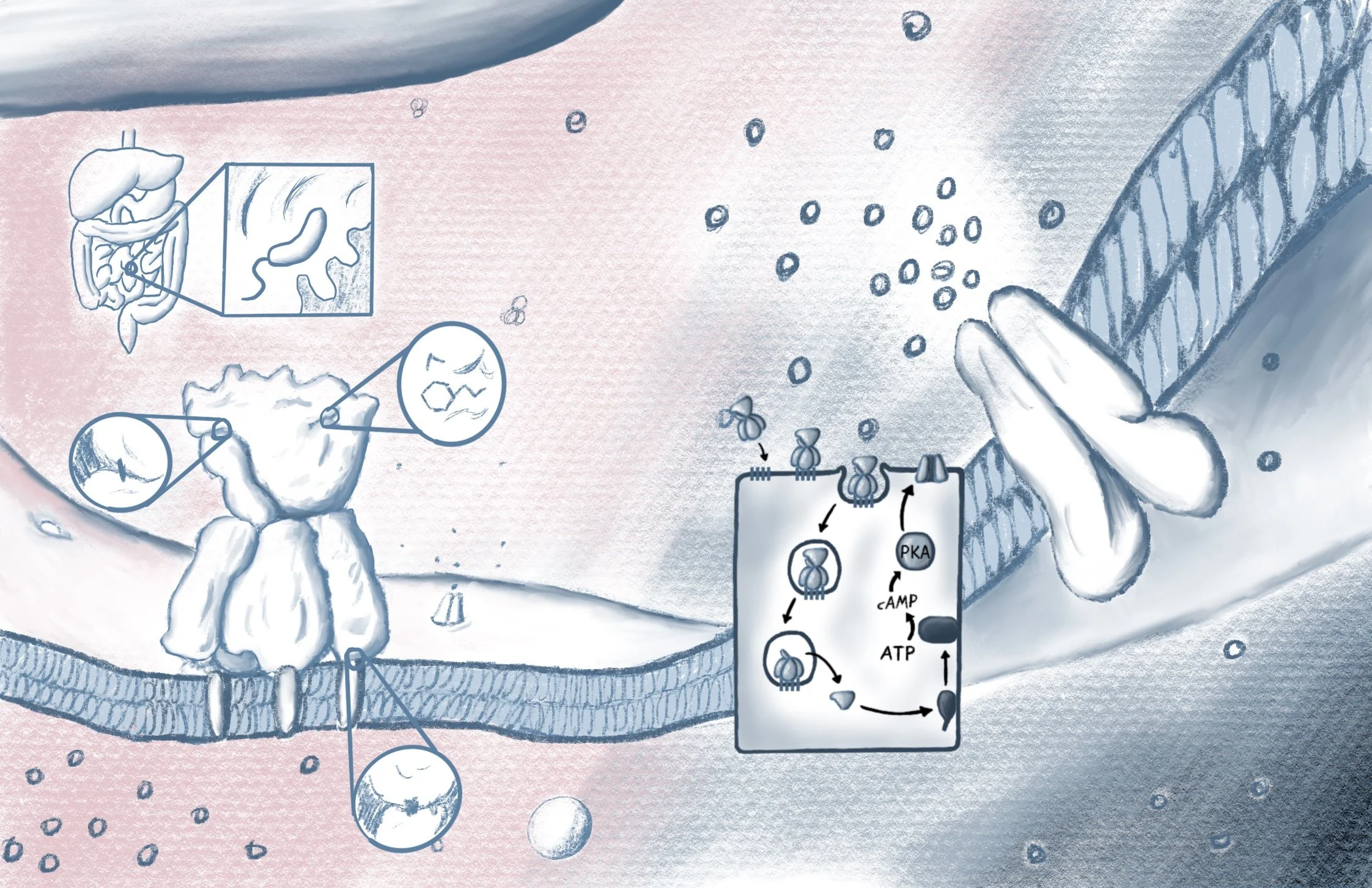
LAM is often not diagnosed until lung collapse - termed pneumothorax - occurs. This is due to it’s rarity and similarity to other diseases, so it is often misdiagnosed first. A major goal of this project was to raise awareness with stunning and engaging visuals, showing the devastating impact of the disease on the lungs. To tell that story, I had the healthy lung on the left and diseased on the right to serve as a direct comparison. Originally I was going to have the right lung collapsed, but decided to instead visualize it in the bottom schematic to complete the story from hypothesized disease mechanism to what usually triggers a diagnosis - pneumothorax.
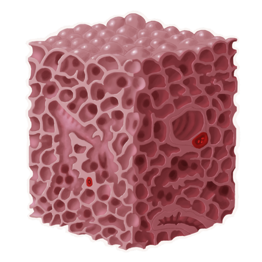
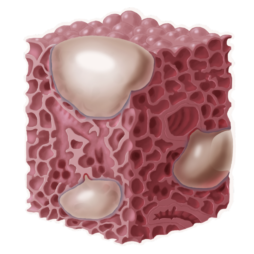
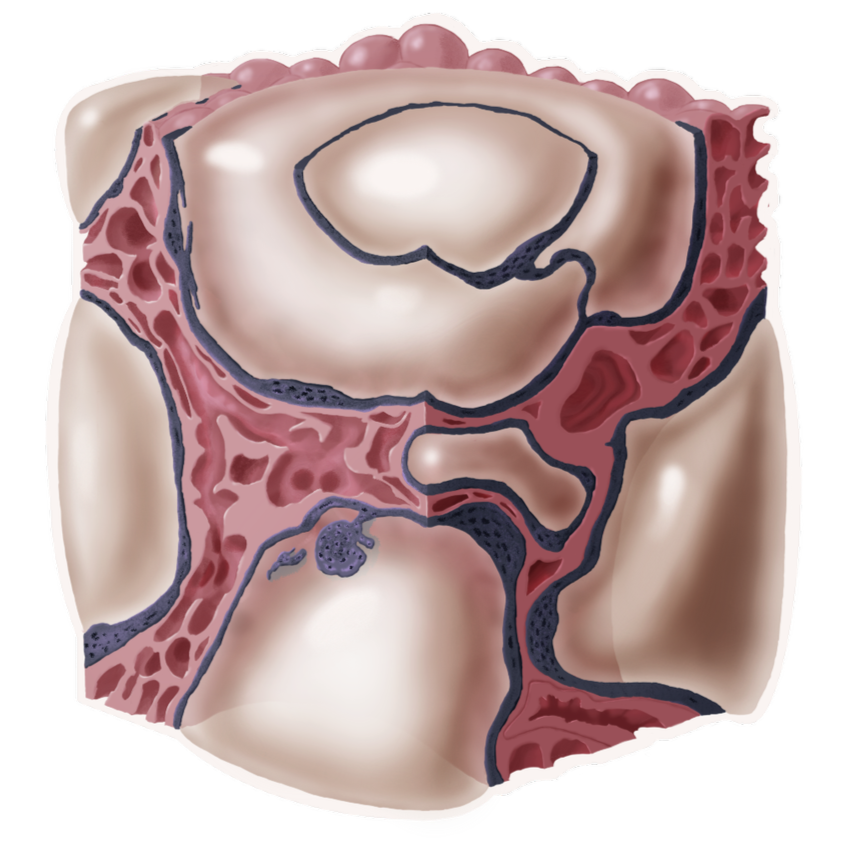
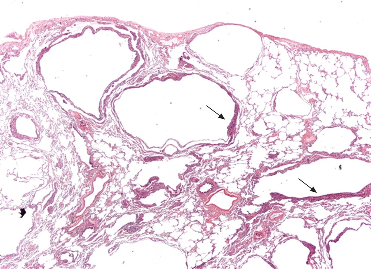
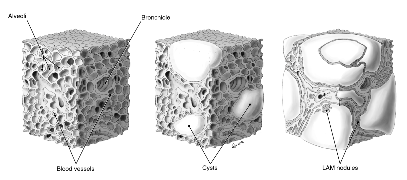
Tissue cubes (left) based on analysis of real histology slides (right) - B. Lazar, 2024.
LAM cell buildup (LAM nodules) is marked by the arrows in the right histology slide. It is theorized that LAM cells travel in the lymph vessels up to the lungs, becoming stuck and embedded at the alveoli. From there, cysts grow until eventually the entire lung is replaced by LAM tissue and cysts.
Other Projects & Gallery
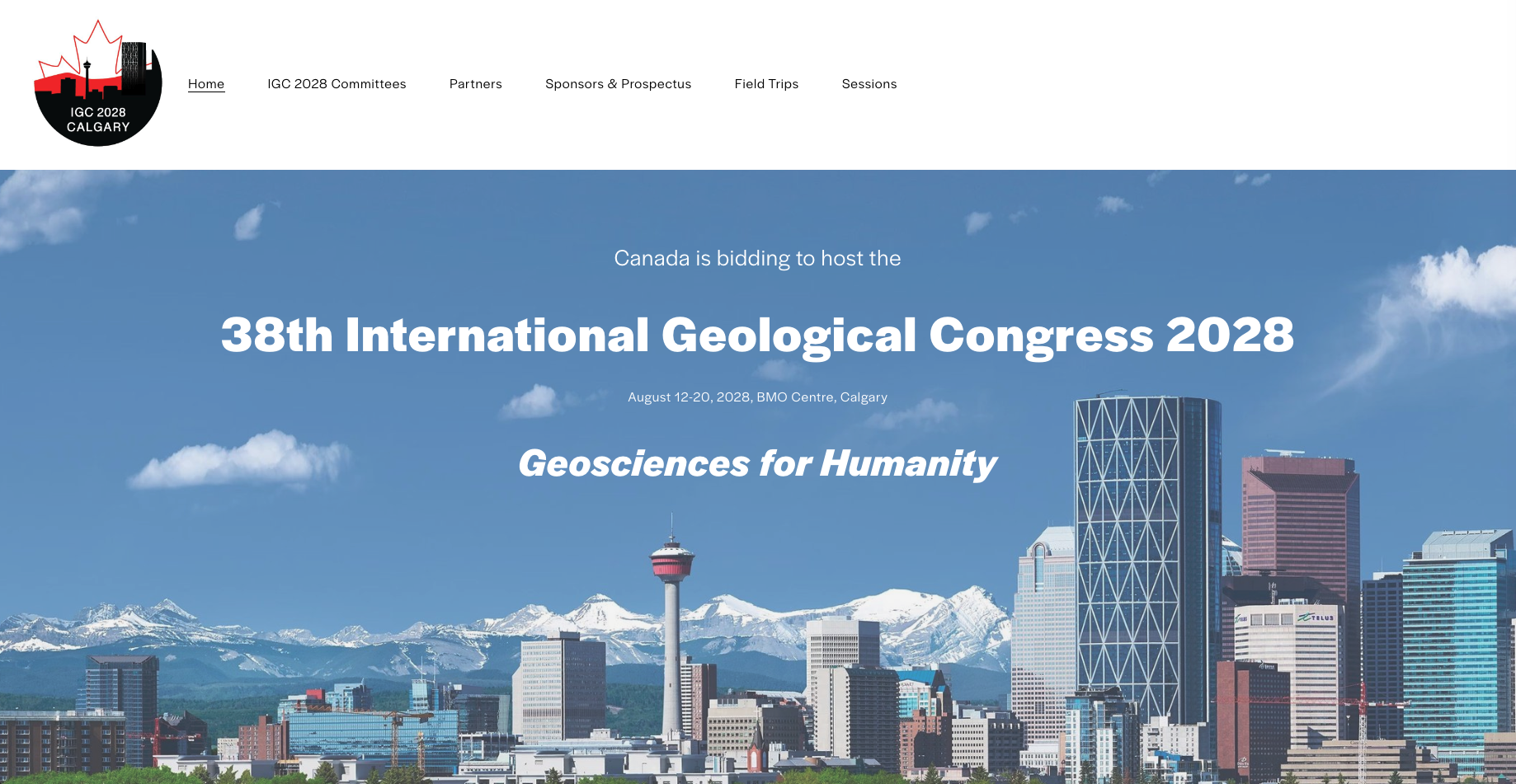
Laying the Foundations for IGC 2028
In 2023, I was contacted to help design the public-facing communications for the bid to host the world’s largest geoscience conference in Calgary for 2028. Through these designs and collaboration with 40+ geoscientists, we won the bid in 2024. Click below to view some of the process work.

Proposed logo design - B.Lazar & IGC Committee, October 2023
Initial website design - B.Lazar & Bid Committee, August 2023

Finalized design for the bid - B.Lazar & Bid Committee, January 2024
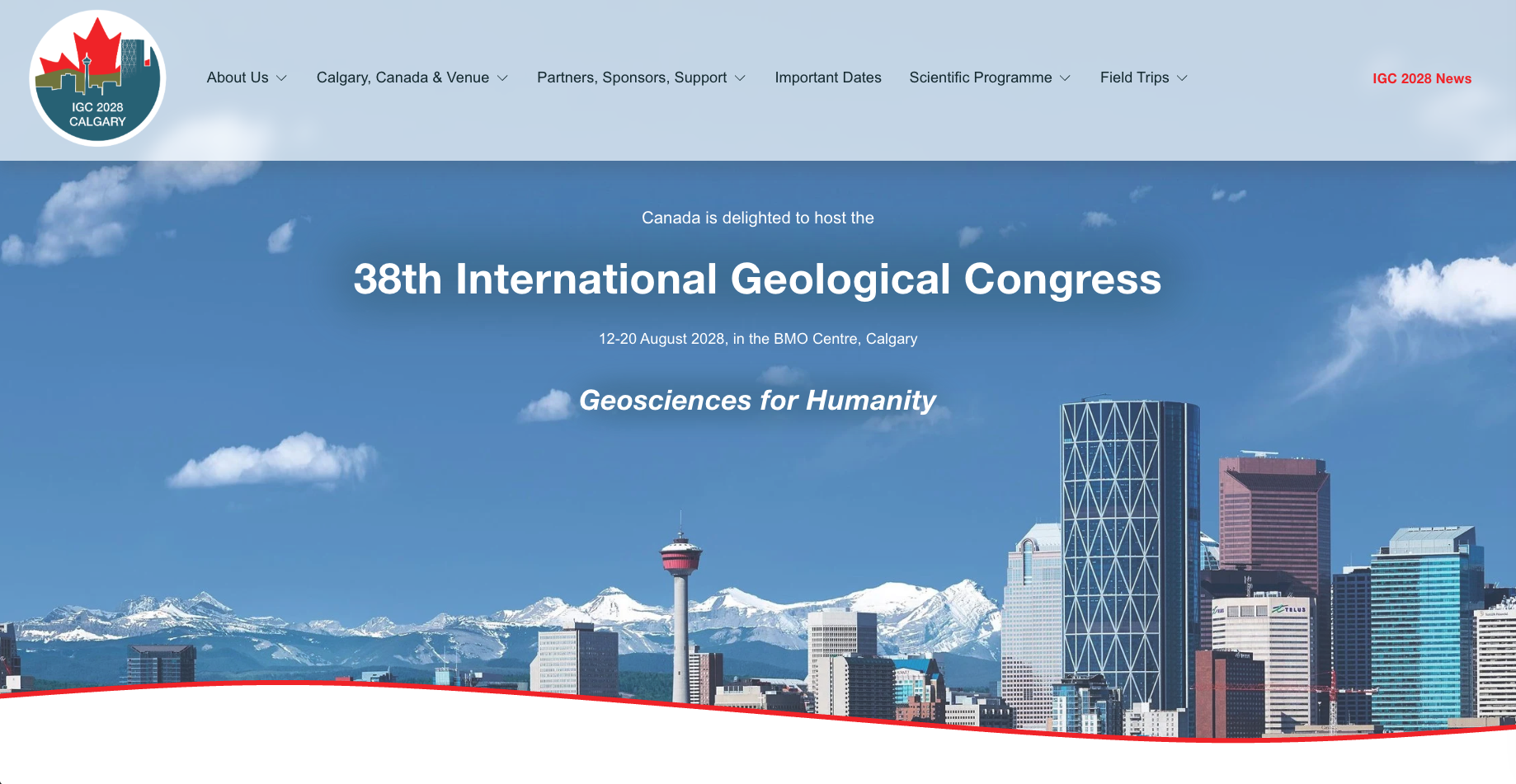
Current design - B.Lazar & IGC Committee, May 2026
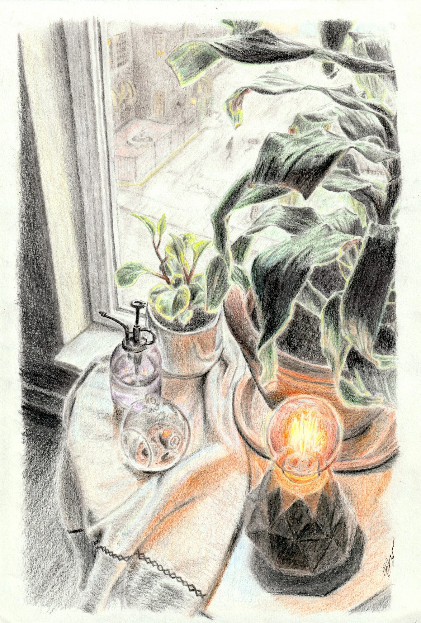
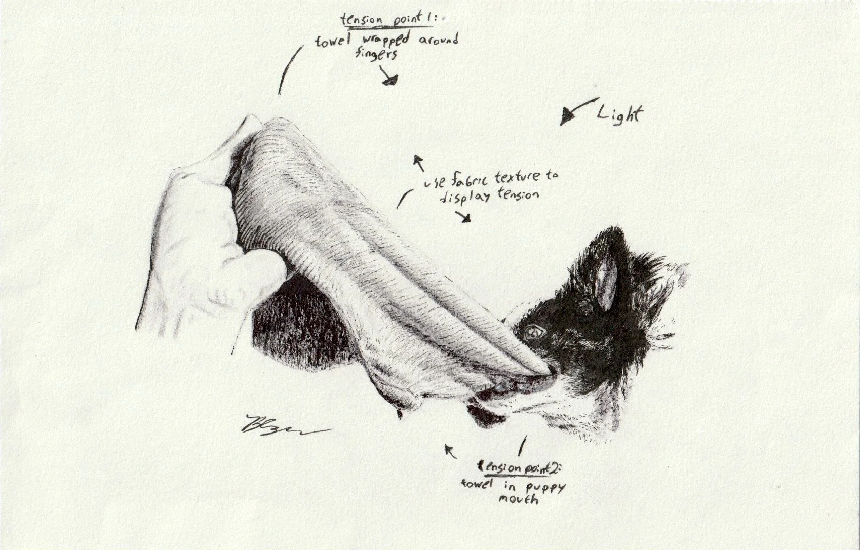
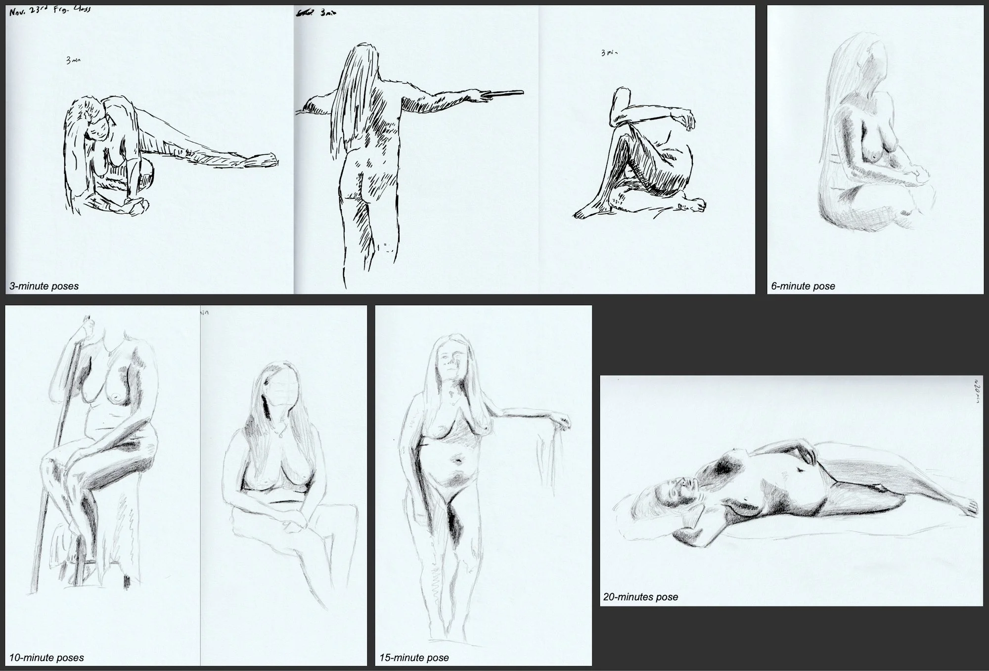
Gallery


Explore More
Learn about my story, background, and career-forming projects
Communicating science with narrative through video, writing, and graphic platforms